Ask in plain language
No PyMOL commands, no installs. You describe the structure and the figure you want; RBAi fetches the data and renders it.
RBAi Academy
A hands-on course in reading real protein structures — using nothing but plain-language prompts in RBAi. Each lesson takes one famous molecule, builds a publication-quality figure, and teaches one big idea you keep for good.
Free to try · no install · real PDB structures
Module 1
From reading your first structure to docking a drug and testing a mutation. Each lesson builds on the one before, and each ends with you doing real research in RBAi.

Open a real protein structure in RBAi, highlight the residues that matter, and get a clean, labelled figure — using plain-language prompts, no code and no software to install.
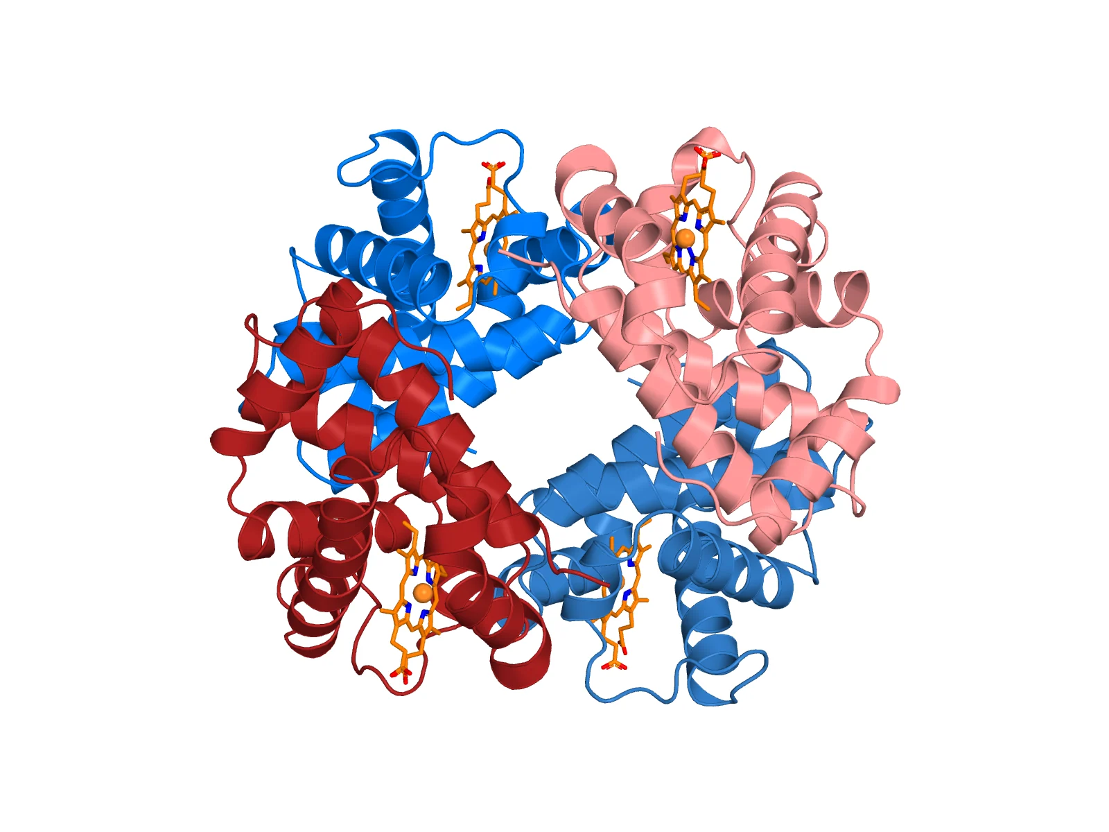
Start lessonOpen hemoglobin in RBAi, see how four protein chains assemble into one oxygen carrier, and zoom to the iron atom a single histidine holds in place — all from plain-language prompts.
Start lesson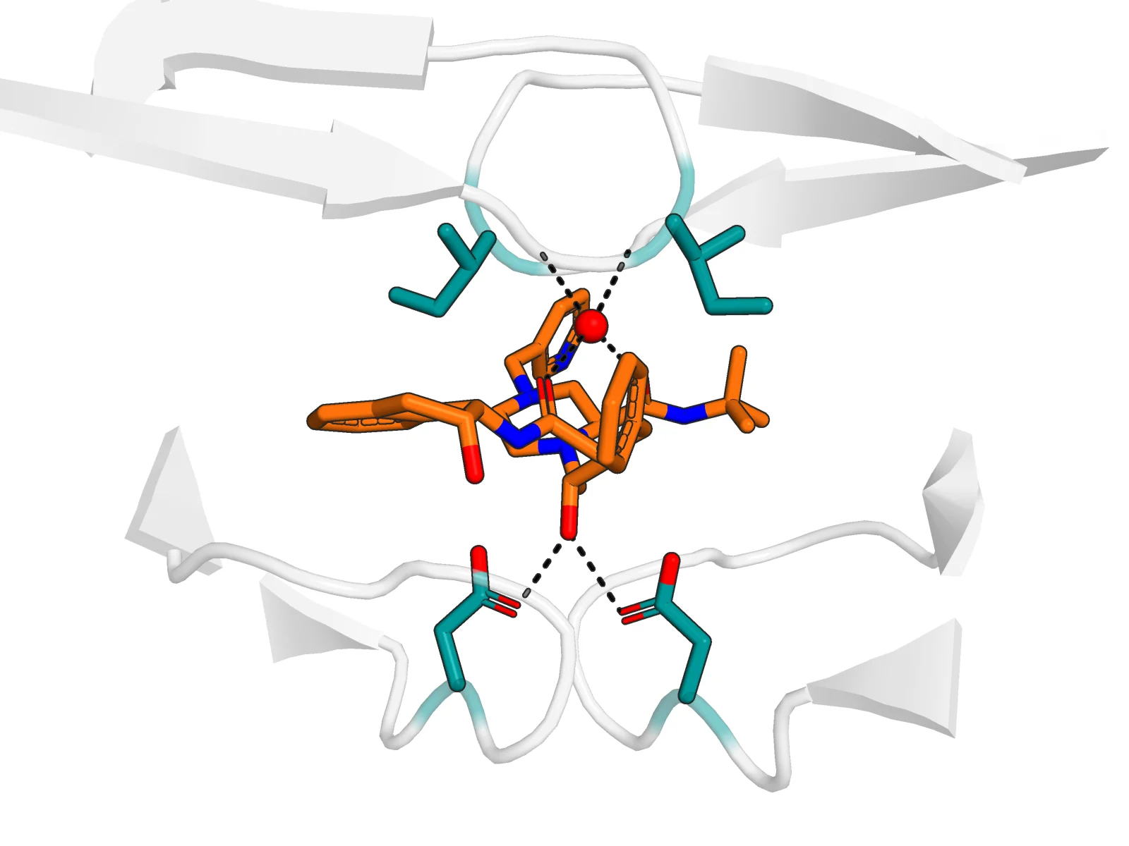
Open HIV-1 protease with the drug indinavir, find every hydrogen bond and hydrophobic contact, measure the catalytic distances, and see how a transition-state mimic jams the enzyme.
Start lesson
Find what the cancer hotspot Arg273 of p53 touches on DNA, mutate it to histidine, find the interactions again, and relax the mutant with an energy minimisation.
Start lesson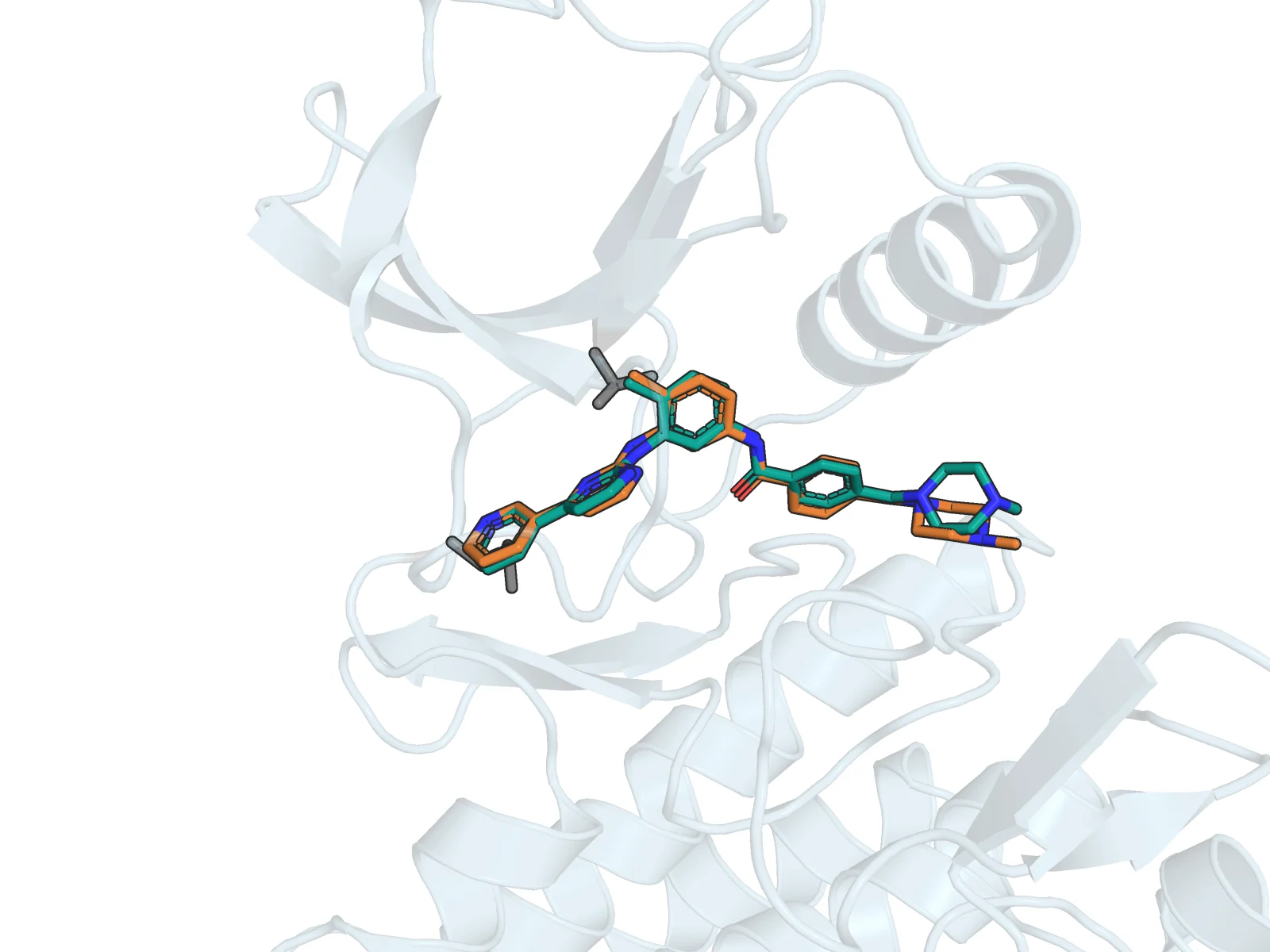
Redock the leukaemia drug imatinib into ABL kinase with AutoDock Vina in RBAi, then check the prediction against the crystal structure. In our run the top pose landed 0.85 Å from the experiment.
Start lesson
Superpose the open and closed structures of adenylate kinase in RBAi, watch two domains swing shut over an inhibitor, and learn why an RMSD only means something once you say which atoms you aligned.
Start lessonThe Journal
What RBAi can do, how to choose between our tools, and how we check the evidence behind them. New articles land here alongside the lessons.
How the Academy works
No PyMOL commands, no installs. You describe the structure and the figure you want; RBAi fetches the data and renders it.
Every lesson teaches you to check the result against the real structure — and to ask for the specific fix when it isn't right yet.
Real PDB structures, real sources, and honest limits on every figure. You learn what a picture can show — and what it can't.
Start with Lesson 1, or jump straight into RBAi and bring a structure of your own.