Dock a cancer drug and check it against the experiment: imatinib in ABL kinase
Redock the leukaemia drug imatinib into ABL kinase with AutoDock Vina in RBAi, then check the prediction against the crystal structure. In our run the top pose landed 0.85 Å from the experiment.
Before 2001, chronic myeloid leukaemia (CML) was usually fatal within a few years. The disease is driven by one runaway enzyme, the BCR-ABL kinase (a kinase is an enzyme that switches other proteins on by adding phosphate groups). Imatinib (Gleevec) blocks that enzyme, and it turned CML into a disease most patients live with. It was one of the first cancer drugs designed against a single protein, and the crystal structure (the 3D atomic model solved from a protein crystal) of imatinib bound to ABL showed how it works: the drug locks the kinase in an inactive shape.
In this lesson you will do what drug designers do before they trust a computer: dock imatinib into ABL (predict, by computer, where and how it sits in the protein) with AutoDock Vina, a widely used docking program, then check the prediction against the experiment. That check is called redocking. You take a drug out of a crystal structure, dock it back in, and ask whether the program finds the pose (the drug's exact position, orientation and shape) the crystal shows. If it can't do that, you shouldn't trust it on a molecule nobody has crystallised.
We use PDB 1IEP (entry 1IEP in the Protein Data Bank, the public archive of solved 3D structures): the ABL kinase domain (the folded region of ABL that does the kinase work) with imatinib (residue name STI, the drug's three-letter code in the file), solved by X-ray diffraction at 2.1 Å by Nagar and colleagues (2002). X-ray diffraction works out atom positions from how a protein crystal scatters X-rays; 2.1 Å is the resolution, the finest detail the data can show (smaller is sharper; one angstrom, Å, is 0.1 nanometres, about the width of an atom).
What you will make

The target. The ABL kinase domain (chain A of PDB 1IEP, one of two protein copies in the file, light blue) has a small N-lobe built from β-strands (top; it comes from the start, or N-terminus, of the chain) and a larger, mostly helical C-lobe (bottom; from the C-terminal end). Imatinib (orange) lies in the cleft between them, where ATP would normally bind, and reaches deep into a pocket that only opens in the inactive kinase.
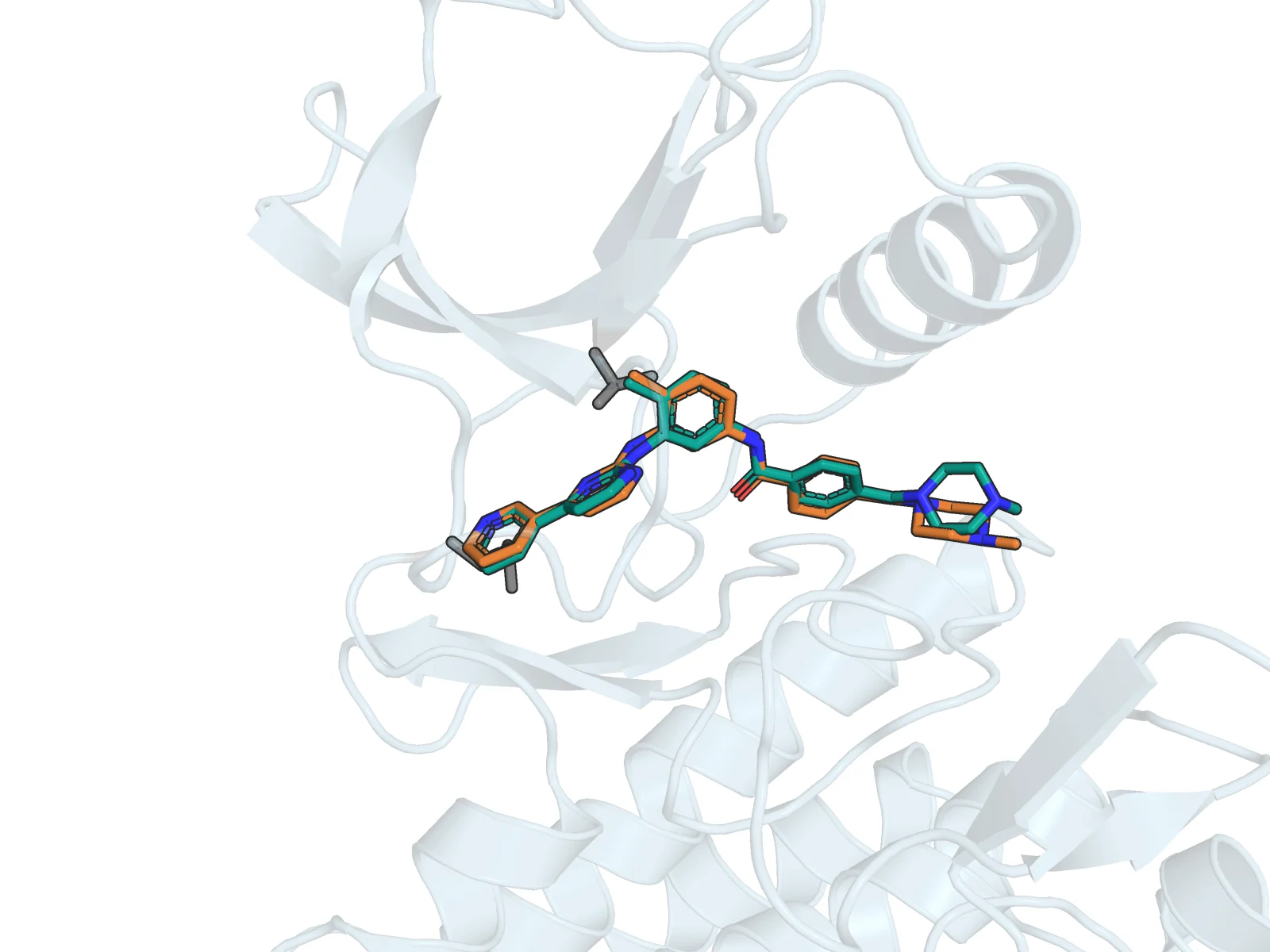
The check. Crystal imatinib (orange carbons) and the top-scoring docked pose from AutoDock Vina (teal carbons) almost coincide: heavy-atom RMSD 0.85 Å over all 37 atoms, in our run (RMSD, root-mean-square deviation, is a kind of average distance between matching atoms; heavy atoms are all atoms except hydrogen). The grey sticks (bonds drawn as rods) are the gatekeeper Thr315 (threonine 315, which guards a pocket at the back of the site; upper left of the drug) and the hinge residue Met318 (methionine 318, in the hinge that joins the two lobes; lower left, next to the pyridine ring). The cartoon (the ribbon drawing of the protein chain) is faded so the drug stays readable. Only the piperazine ring at the right end differs visibly.
1. Open the structure Main chat
Start a new chat in RBAi:
Open PDB 1IEP as an interactive 3D structure. Tell me the chains, the ligand residue name and number of imatinib in each chain, method and resolution. Do not change the structure.
What you should see: a 3D card, plus two chains (A and B). Each chain is one ABL kinase domain with its own imatinib, STI 201 in chain A and STI 202 in chain B. The ligand is the small molecule bound to the protein (here imatinib), and the file numbers it like a residue, an amino acid of the chain. The method is X-ray diffraction at 2.1 Å. Two copies of the drug matter for everything that follows: always say which one you mean.
2. Make the overview figure Edit with AI
Click Edit with AI on the 3D card. This opens Structure Studio, where RBAi works on the structure in PyMOL, a standard molecular-graphics program.
Make a figure of the whole chain A kinase domain as an opaque light blue cartoon with only the crystal imatinib as thicker orange sticks. N-lobe at the top, C-lobe at the bottom, imatinib lying across the cleft between them. White background, nothing clipped.
What you should see: the first figure above, a ray-traced (realistically lit) 1600×1200 image in the studio. Our first render showed the kinase from the back with the drug hidden behind helices, so we asked once more for this orientation.
3. Redock imatinib Edit with AI
Redock imatinib to check the docking setup: dock the crystal ligand resn STI and chain A back into chain A only (exclude chain B), with the box drawn around resn STI and chain A and a 3 Å margin.
RBAi starts AutoDock Vina as a background run. (In the prompt, resn STI selects atoms by residue name, and the box is the region of space Vina searches: here the crystal imatinib of chain A plus a 3 Å margin.) It removes the crystal ligand and waters from the receptor (the protein the drug docks into), rebuilds imatinib from its chemical definition and searches the box for the best poses. When the run finishes, use Show poses in Edit with AI to step through them. Pose 1, together with the receptor, becomes a new version of the structure (Edit with AI saves each change as a numbered version; version 0 is the original crystal structure).
What you should see: in our run the box-and-search took a few minutes and returned 7 poses, scored from −12.4 (pose 1) to −9.5 kcal/mol (more negative means a better predicted fit). Pose 1 touches Glu286, Thr315, Met318, Ile360, His361 and Asp381, which are the pocket residues the crystal ligand touches.
One thing went wrong first, and it's worth knowing. Our first attempt selected the ligand as just resn STI. That picks up both imatinib copies, so the box covered both chains' sites, more than 27,000 ų. Vina's best poses then sat 10 to 19 Šaway from the crystal pose. Restricting the selection to resn STI and chain A fixed it. A docking box is a hypothesis about where the drug binds, so check what it covers.
4. Compare pose 1 with the experiment Edit with AI
Check this redocked pose 1 against the experiment. Superpose version 0 (the crystal structure) onto this version so the crystal imatinib of chain A comes in next to the docked one. Measure, with PyMOL and without fitting, the heavy-atom RMSD between docked and crystal imatinib. Atom names differ after docking, so pair atoms by element with an optimal one-to-one assignment, not by name.
Superposing lays one structure on top of another so they can be compared atom by atom. What you should see: in our run, RMSD 0.85 Å over 37 heavy atoms. That is well under the usual 2 Å threshold, so the setup reproduces the experiment. The "without fitting" part matters: the receptor never moved, so the number measures where Vina put the drug, not how well two drugs can be overlaid.
Asking for element-based pairing also matters. A docked ligand comes back with new atom names. When we first let PyMOL pair atoms by name, only 24 of 37 atoms matched and the number (13 Å) was meaningless. We checked the 0.85 Å independently from the downloaded coordinates, and both drugs' centres lie within 0.4 Å of each other.
5. Make the comparison figure Edit with AI
Show only this receptor as a light blue cartoon, hide the overlay protein, show the crystal imatinib with orange carbons and the docked imatinib with teal carbons as sticks, and Thr315 and Met318 of chain A as grey sticks. Zoom on the two imatinib copies plus Thr315 and Met318 with about 4 Å of margin, rotate so imatinib lies flat across the picture, make the cartoon 70% transparent (set cartoon_transparency, 0.7 and set stick_radius, 0.22). White background, no labels.
That is the second figure above. Our first version used an opaque cartoon that crossed in front of the drug, so we added the transparency in a second request. The two set commands in brackets are PyMOL settings that pin our exact values; describing the look in words also works.
The ideas behind the picture
- Kinases switch shape. A loop with the motif Asp-Phe-Gly (DFG) flips between an "in" (active) and an "out" (inactive) position. Imatinib binds the DFG-out form. Its long body runs from the hinge (Met318), past the gatekeeper (Thr315), into a back pocket that only exists when Asp381-Phe382 has flipped out. Binding the inactive shape is part of why imatinib is selective.
- Docking = search + score. Vina tries many placements and conformations (shapes the flexible drug can bend into) of the drug inside the box (search) and ranks them with an empirical function, a formula fitted to experimental data (score). A pose can be wrong because the search never found the right one, or because the score ranked a wrong one first.
- Redocking tests the setup. If the program can put a known drug back where the crystal shows it (heavy-atom RMSD under about 2 Å), your box, receptor and ligand preparation are reasonable. Only then is it worth docking new molecules.
- RMSD needs the right atom pairs. RMSD is the root-mean-square distance between matched atoms. Matching them wrongly, by atom names that changed, gives a wrong number with full confidence.
What the picture shows, and what it doesn't
- A Vina score of −12.4 kcal/mol is an estimate from a scoring function, not a measured binding affinity. Use scores to rank poses within one run, not to say how strongly a drug binds.
- Redocking into the crystal receptor is the easy case. The protein already has the shape it took around imatinib. Docking into a structure solved without the drug, or into a different kinase, is much harder, and Vina keeps the protein rigid.
- The figure shows one chain, with no waters and no protein motion. It is a static snapshot of one crystal, not the drug's whole binding pathway.
Your turn
In patients, the mutation T315I (threonine 315 to isoleucine) makes CML resistant to imatinib. Look at the close-up: Thr315 sits right against the middle of the drug, next to the amine that links imatinib's two halves. Before you ask RBAi anything, write down two reasons why swapping threonine (small, with an OH group) for isoleucine (bulkier, no OH) could stop imatinib binding. Then ask RBAi in Edit with AI to measure the distances from the Thr315 side chain to imatinib, and check your reasoning. Bonus, as a researcher: if you mutated Thr315 to Ile and redocked, what result would support your idea, and what would not? Remember that Vina keeps the mutant protein rigid.
Try it
Open RBAi and run the redocking yourself, starting with step 1. If your RMSD comes out large, check the box selection first, then the atom pairing.