Inside an active site: how a drug blocks HIV-1 protease
Open HIV-1 protease with the drug indinavir, find every hydrogen bond and hydrophobic contact, measure the catalytic distances, and see how a transition-state mimic jams the enzyme.
HIV cannot make new infectious virus without one small enzyme: HIV-1 protease. It cuts the virus's long protein chains into working parts. Block it, and the virus particles stay immature and cannot infect. Indinavir, approved in 1996, was one of the protease inhibitors that turned HIV from a death sentence into a manageable disease, and it was designed with crystal structures (3D atomic models solved from protein crystals) exactly like the one in this lesson.
In Lessons 1 and 2 you looked at structures. Here you start to analyse one, the way a medicinal chemist does: find every hydrogen bond (a weak attraction between a hydrogen on one nitrogen or oxygen atom and another N or O nearby) between the drug and the enzyme, see which oily, water-avoiding (hydrophobic) side chains (amino-acid R groups) wrap around it, and measure the distances that explain why it binds. You do the analysis in Edit with AI (Structure Studio, opened with the Edit with AI button on the 3D structure card), RBAi's workspace that runs PyMOL, a standard molecular-graphics program, on your structure. You make the figures in the main chat.
We will use PDB 1HSG (entry 1HSG in the Protein Data Bank, the public archive of solved 3D structures): HIV-1 protease with indinavir bound, solved by X-ray diffraction (2.0 Å as listed by RCSB, the Protein Data Bank's US site) by Chen and colleagues at Merck in 1994. X-ray diffraction works out atom positions from how a protein crystal scatters X-rays; 2.0 Å is the resolution, the finest detail the data can show (smaller is sharper; one angstrom, Å, is 0.1 nanometres, about the width of an atom). In the file the drug is called MK1.
What you will make
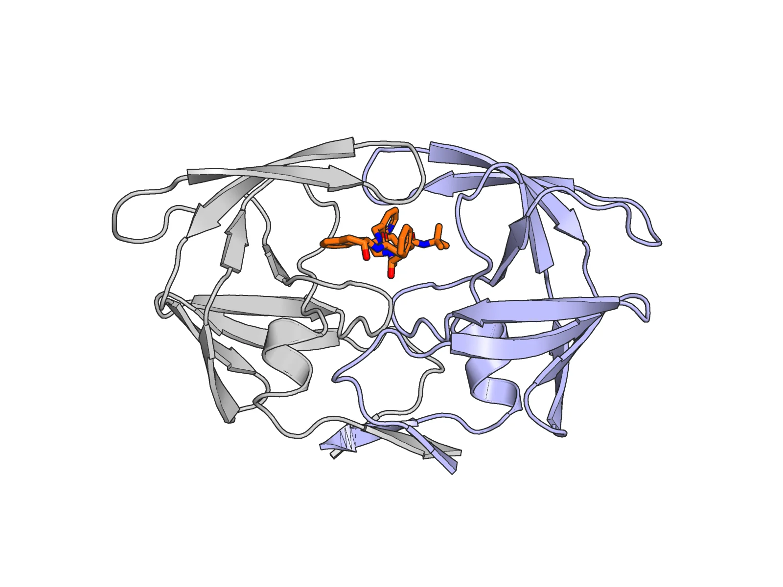
The whole enzyme. HIV-1 protease is two identical protein chains (together, a dimer), A (light blue) and B (light grey), drawn as cartoon: ribbons that trace each chain. Together they build one tunnel at the interface, and indinavir (orange sticks, every bond drawn as a rod) sits inside it. The two β-hairpin flaps (each two strands joined by a tight turn) at the top have closed down over the drug. PDB 1HSG.
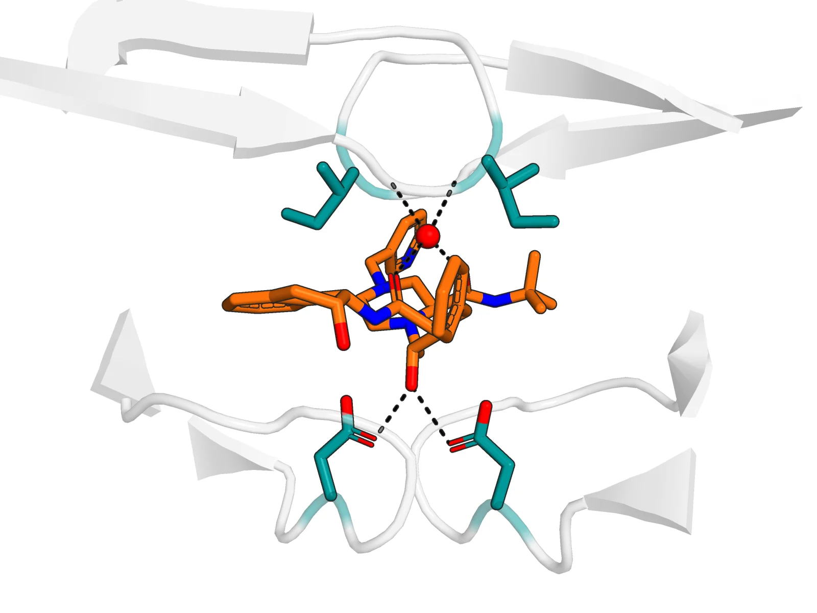
The active site, same orientation. At the bottom, the catalytic Asp25 (chain A) and Asp25′ (chain B), the aspartic acid at position 25 of each chain (three-letter amino-acid code plus position), both reach the drug's central hydroxyl, its OH group (dashed lines). At the top, the flap residues (amino acids) Ile50 and Ile50′ (isoleucine 50 of each chain, teal) hold one water (HOH 308, water number 308 in the file, red sphere) that hydrogen-bonds to two carbonyl (C=O) oxygens of indinavir. One drug, gripped from below by the chemistry and from above by the flaps.
1. Open the structure Main chat
Start a new chat in RBAi:
Open PDB 1HSG as an interactive 3D structure. Tell me the chains, the ligand residue name and number, and whether there are crystallographic waters near the ligand. Do not change the structure.
What you should see: a 3D card you can rotate, two protein chains A and B (99 residues each), and the drug as MK1, residue 902 of chain B. The ligand is the small molecule bound to the protein, here the drug, and the file numbers it like a residue. In our run RBAi also found 9 waters within 4 Å of the drug, the closest at 2.68 Å. These are crystallographic waters: water molecules held still enough in the crystal to be seen in the model. Keep those waters in mind; one of them matters.
2. Make the overview figure Main chat
Still in the main chat:
Make a publication-quality overview figure of PDB 1HSG. White background, remove solvent, both chains as cartoon, chain A light blue and chain B soft light grey, indinavir (resn MK1) as orange sticks with heteroatoms coloured by element, sitting in the tunnel at the dimer interface. Use the PyMOL figure renderer.
RBAi writes the PyMOL scene (the instructions that set up the picture) and returns a ray-traced image, rendered with realistic light and shadow. Heteroatoms coloured by element means atoms other than carbon get their standard colours, such as red for oxygen and blue for nitrogen. Our first render came out upside down (flaps at the bottom) with a lot of empty space, so it took a few follow-ups (the same scene turned 180° and zoomed tighter) to get the first figure above. Find the flaps yourself: they are the two hairpins that cross over the drug.
3. Find the hydrogen bonds Edit with AI
On the 3D structure card, click Edit with AI and ask:
Find the hydrogen bonds between indinavir (resn MK1) and the protease, and any water that bridges the drug to the protein. List each one with the residue, chain, atom names and distance.
What you should see: dashed lines drawn on the structure and a list. In our run RBAi found six direct hydrogen bonds (N/O pairs within 3.5 Å that pass PyMOL's geometry test). Atoms are listed by their names in the file (O2 is one of indinavir's oxygens; OD1 is one of the two oxygens at the tip of the Asp side chain), and residues by code and number (Gly27 is glycine 27, Arg8 is arginine 8):
- indinavir O2 to Asp25 OD1 of chain B, 2.63 Å, and of chain A, 2.77 Å
- indinavir N4 and O4 to the Gly27 backbone oxygen of chain B (3.03 and 3.16 Å)
- O4 to the Asp29 backbone nitrogen of chain B (3.23 Å)
- N5 to Arg8 of chain B (3.46 Å)
It also found two bridging waters. The important one is HOH 308: it touches indinavir's O1 (2.68 Å) and O3 (2.80 Å) and the backbone nitrogens of Ile50 in chain A (3.11 Å) and chain B (2.93 Å). That is the famous flap water, a single water molecule tying both flaps to the drug.
4. Find the hydrophobic contacts Edit with AI
Now find the hydrophobic contacts between indinavir and the protease, and list the residues that line the pocket around it (within 4 Å), by chain.
What you should see: in our run, 13 hydrophobic contacts (carbon pairs within 4.0 Å), the closest from Val32 of chain B (3.32 Å). The pocket is lined by the same residues from both chains: Arg8, Leu23 (A), Asp25, Gly27, Ala28, Asp29/Asp30 (B), Val32, Ile47, Gly48, Gly49, Ile50 (B), Pro81, Val82 and Ile84. Notice how many are oily (Val, Ile, Leu, Pro, Ala: valine, isoleucine, leucine, proline, alanine). Hydrogen bonds aim the drug; hydrophobic contacts hold most of its bulk.
5. Measure the catalytic distances Edit with AI
Measure the distance from indinavir's central hydroxyl oxygen to both carboxylate oxygens of Asp25 in chain A and of Asp25 in chain B, and draw them as dashed lines.
What you should see: in our run the central hydroxyl is O2, and its distances are 2.77 and 2.93 Å to Asp25 OD1/OD2 of chain A (the two oxygens of the carboxylate, the COO⁻ group at the tip of the side chain) and 2.63 and 3.04 Å to Asp25 OD1/OD2 of chain B. All four are hydrogen-bonding distances: the hydroxyl sits right between the two catalytic acids.
6. Make the close-up figure Main chat
Same as the overview, you only describe the picture you want. In the main chat:
Make a publication-quality close-up of the indinavir (resn MK1) binding site in PDB 1HSG. Show the drug as orange sticks. Show the two catalytic Asp25 residues and the two flap Ile50 residues (chains A and B) as teal sticks, and keep the surrounding active-site loops as a faint, semi-transparent grey cartoon. Show the flap water HOH 308 as a small red sphere. Draw dashed distance lines from the drug's O2 to each Asp25 OD1, from the drug's O1 and O3 to the water, and from the water to each Ile50 backbone nitrogen. White background, heteroatoms coloured by element, and zoom in tight on the drug, those residues and the water. Use the PyMOL figure renderer.
Every residue and atom in that prompt is one you already found in steps 3 and 5, so RBAi has all it needs to build the scene. That is the second figure above.
Optional — reproduce it exactly. You never have to write PyMOL. But if you want a pixel-for-pixel copy of our figure instead of RBAi's own framing, you can also paste the exact scene below into the main chat and ask RBAi to render it with the PyMOL figure renderer (Edit with AI can run it too), because both run PyMOL for you. We used this one:
cmd.select("site", "chain A+B and resi 25+50 and polymer.protein")
cmd.select("wat", "resn HOH and resi 308")
cmd.show("cartoon", "polymer.protein and resi 22-32+44-56")
cmd.color("grey90", "polymer.protein and not site")
cmd.set("cartoon_transparency", 0.5)
cmd.show("sticks", "resn MK1")      # indinavir, orange carbons
cmd.show("sticks", "site")          # Asp25/25' and Ile50/50', teal carbons
cmd.show("spheres", "wat")          # the flap water, small red sphere
cmd.set("sphere_scale", 0.3, "wat")
cmd.distance("hb", "resn MK1 and name O2", "chain A and resi 25 and name OD1")
cmd.distance("hb", "resn MK1 and name O2", "chain B and resi 25 and name OD1")
cmd.distance("hb", "resn MK1 and name O1", "wat")
cmd.distance("hb", "resn MK1 and name O3", "wat")
cmd.distance("hb", "wat", "chain A and resi 50 and name N")
cmd.distance("hb", "wat", "chain B and resi 50 and name N")
cmd.hide("labels", "hb")
cmd.orient("polymer.protein or resn MK1")
cmd.turn("z", 180)
cmd.zoom("resn MK1 or site or wat", buffer=2.5)
Every atom name in it (O1, O2, O3, OD1, N) is one you reported in steps 3 and 5, so each dashed line is a contact you measured yourself. (The scene also sets the colours, stick and dash sizes and a white background.) The plain prompt above and this script give you an equivalent figure; the script just pins every colour, distance and angle to match ours exactly. Describing what you want in words is enough.
The ideas behind the picture
- One active site from two chains. Each chain brings one Asp25. Only the dimer has the pair, so only the dimer can cut. That is why the drug sits exactly on the two-fold axis.
- Catalysis by two aspartates. In the real reaction, the two Asp25 carboxylates activate a water that attacks the peptide bond, passing through a tetrahedral, hydroxyl-bearing intermediate (a short-lived state in which the attacked carbon carries four groups, one of them an OH).
- A transition-state mimic. The transition state is the reaction's short-lived, highest-energy peak, and a mimic is a stable molecule shaped like it. Indinavir's central hydroxyl sits where that intermediate's hydroxyl would sit, between both aspartates (step 5). But its hydroxyethylene core (a –CH(OH)–CH₂– link where a substrate would have its peptide bond) has no bond the enzyme can cut. The enzyme grips it as if it were the reaction's peak and cannot finish the job.
- Flaps and the flap water. The flaps close over the drug, and in 1HSG a single water (HOH 308) links both Ile50 backbone nitrogens to two drug carbonyls. Many inhibitors are designed around that water, and some later drugs were designed to replace it.
- Reading contacts. Hydrogen bonds (about 2.6 to 3.5 Å between N and O) are directional and set the drug's position. Hydrophobic contacts (carbon to carbon, up to about 4 Å) are weak one by one but numerous.
What the picture shows, and what it doesn't
- What you can point to: which atoms are close enough to hydrogen-bond, which side chains pack against the drug, and where the flap water sits. Those are measurements on the deposited coordinates.
- What depends on the rules: "hydrogen bond" and "hydrophobic contact" are distance and geometry cutoffs. Change the cutoff and the count changes. X-ray structures also have no hydrogen atoms, so donors and acceptors (which atom gives the hydrogen and which receives it) are inferred.
- What it cannot tell you: binding strength, how the flaps open and close in solution, or how the drug behaves in a patient. A crystal structure is one well-ordered snapshot, and the protonation of the two aspartates (which one carries an extra hydrogen) is not visible in it.
Your turn
You now know which residues line the pocket. HIV mutates fast, and resistance often comes from a single change in that lining. Two classic indinavir-resistance mutations are V82A (valine 82 changed to alanine, written in one-letter amino-acid codes) and I84V (isoleucine 84 changed to valine).
In Edit with AI, find Val82 and Ile84 in your contact list. For each, ask: which drug atoms does it touch, and what does the mutation do to the side chain (V82A loses two carbons; I84V loses one)? Predict which contacts would weaken. Then write one sentence on why a mutation that makes the pocket slightly larger can hurt a drug more than it hurts the virus's own substrates (the viral protein chains it normally cuts). You will make a mutation like this yourself in Lesson 4.
Try it
Open RBAi, paste the first prompt, then click Edit with AI on the 3D structure and run your own analysis. Your numbers should match ours, because the coordinates are the same deposited entry. If a figure needs a different angle, say exactly what to change and look again.