Hemoglobin: four subunits and the iron that carries oxygen
Open hemoglobin in RBAi, see how four protein chains assemble into one oxygen carrier, and zoom to the iron atom a single histidine holds in place — all from plain-language prompts.
Every red blood cell is packed with hemoglobin — the protein that picks up oxygen in your lungs and drops it off in your tissues. It is one of the most studied molecules in biology, and it is a perfect second lesson, because it is not one protein chain (one folded string of amino acids) but four, working as a single machine. That idea — several chains assembling into one functional unit — is called quaternary structure, and hemoglobin is its textbook example.
In Lesson 1 you read a single chain and a binding site. Here you will open a four-chain assembly (a tetramer), colour it so its architecture is obvious, and then zoom all the way in to the single iron atom that actually grabs the oxygen. Same workflow as before: you ask RBAi in plain language in the RBAi main chat, where every prompt in this lesson goes, and you inspect what comes back.
We will use PDB 2HHB (entry 2HHB in the Protein Data Bank, the public archive of solved 3D structures), human deoxyhaemoglobin (haemoglobin with no oxygen bound) — the classic 1.74 Å structure (one angstrom, Å, is 0.1 nanometres, about the width of an atom) from Fermi, Perutz and colleagues.
What you will make
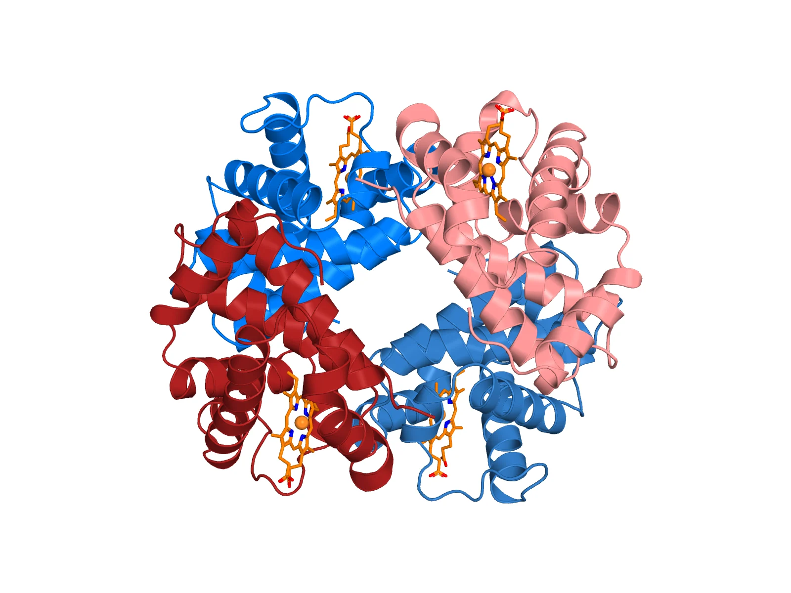
The whole molecule. Four chains, each called a subunit — two α subunits (blue) and two β subunits (red) — pack around a central cavity. That α₂β₂ arrangement is hemoglobin's quaternary structure. Each subunit cradles one orange heme group (a flat, iron-holding ring molecule that is not protein) with an iron atom (sphere) at its centre: four hemes, four irons, four oxygen-binding sites. PDB 2HHB.

One heme pocket, up close. The flat orange ring is the heme; its four nitrogens point inward and hold the iron (sphere). The teal side chain (an amino acid's R group) below the iron is the proximal histidine (His F8: the eighth residue, or amino acid, of helix F) — the single amino acid that anchors the iron to the protein. Oxygen binds on the opposite, open face of the iron.
1. Open the structure
Start a new chat in RBAi:
Open PDB 2HHB and show it as a 3D structure. Tell me how many protein chains it has, which are the alpha and which are the beta subunits, what the heme groups and iron atoms are, and the method and resolution. Do not change the structure.
You should learn that 2HHB has four chains — two α (141 residues) and two β (146 residues) — plus four heme groups, each with one iron, solved by X-ray diffraction at 1.74 Å. X-ray diffraction works out the atom positions from how a protein crystal scatters X-rays, and 1.74 Å is the resolution: the finest detail the data can show, where smaller is sharper.
2. Make the quaternary-structure figure
Make a figure of the whole tetramer: show all four chains as cartoon, colour the two alpha chains blue and the two beta chains red so the α₂β₂ architecture is obvious, and show the four heme groups as orange sticks with their iron atoms as spheres. White background, nothing clipped.
That is the first figure above. Cartoon draws each chain as a ribbon with its helices as coils, sticks draw every bond as a rod, and spheres draw atoms as balls. Now rotate the live 3D structure in the chat: notice the two-fold symmetry (turn it half a circle about its centre and it looks the same), and the cavity at the centre where the four subunits meet. The subunits are not just stuck together — that interface (the surfaces where they touch) is how they communicate.
3. Zoom into one heme pocket
Now zoom into one subunit's heme: fade the rest of that chain to a light cartoon, show the heme as orange sticks with the iron as a sphere, and show the histidine that binds the iron from below — the proximal histidine. Label it.
Look at the second figure. The iron sits in the middle of the flat heme ring, and the teal histidine reaches up to hold it from underneath. That single bond — iron to histidine — is the link between the oxygen chemistry and the whole protein.
4. Ask how it actually carries oxygen
Explain how this structure carries oxygen: what the iron does, why the proximal histidine matters, and what "cooperativity" between the four subunits means. Separate what this deoxy structure actually shows from what comes from textbooks.
The ideas behind the picture
- Quaternary structure: four chains (α₂β₂) form one working molecule. Many proteins only work as assemblies — hemoglobin is the classic case.
- A prosthetic group: the heme is not protein. It is a small molecule the protein holds, and without it hemoglobin cannot carry oxygen. The iron at its centre is where oxygen actually binds.
- One residue does a lot: the proximal histidine is the only direct protein bond to the iron. When oxygen binds on the other side, the iron shifts, the histidine pulls on its helix, and that tiny motion is passed between subunits — cooperativity, which is why hemoglobin loads and unloads oxygen so efficiently.
- Structure meets disease: change one amino acid on the β chain's surface (Glu6→Val: the glutamate at position 6 replaced by valine) and you get sickle-cell haemoglobin. One residue, a different molecule — the reason structural biology matters.
What the picture shows — and what it doesn't
This is deoxyhaemoglobin: no oxygen is bound, so the open face of each iron is empty here. The figure shows the architecture and the iron's protein anchor; it does not show oxygen binding, the motion between states, or binding strengths. Seeing where oxygen would bind is not the same as measuring how tightly it does.
Your turn
In RBAi, rotate the tetramer until you can point to all four irons. Then ask RBAi to show the proximal histidine in a different subunit than the one above, and confirm it is the same residue doing the same job. Quaternary structure means the same part, repeated — find the repeat yourself.
Try it
Open RBAi and build your own hemoglobin figures from the prompts above. As always, ask for a clearer angle or colour and look again — that back-and-forth is how real figures get made.